Klinische Fälle
deformități

Wirbelsäulendeformitäten

Skoliose und Kyphose — idiopathisch, neuromuskulär, syndromal (Larsen, Parkinson). AIS, kombinierte Verfahren.

Fallserie

Kombinierter Zugang bei idiopathischer Skoliose (AIS)

21 Folien
Slide 1

Combined anterior-posterior approach in surgical treatment of idiopathic scoliosis

Ioan Branea

Center for Spine Surgery, Orthopedics, and Traumatology,

SRH Klinikum Karlsbad-Langensteinbach

Karlsbad, Germany

Slide 2

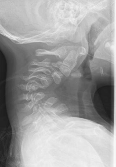
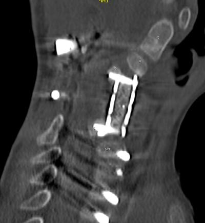
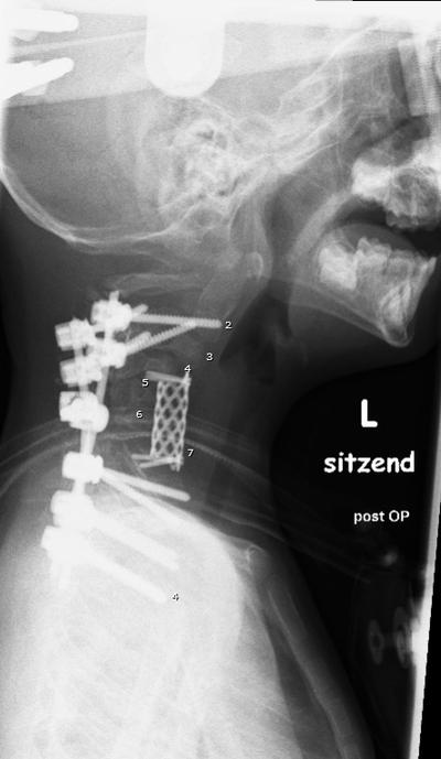
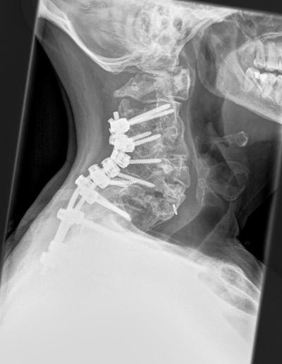
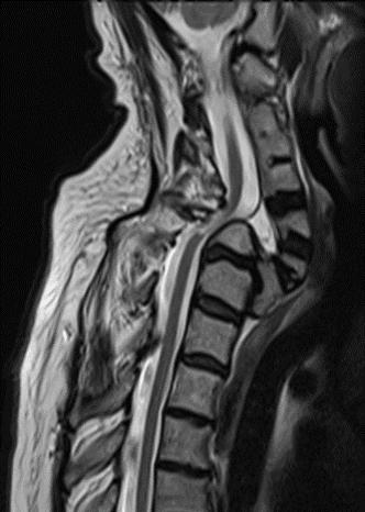
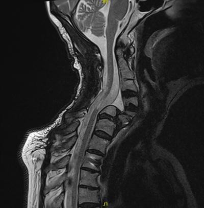
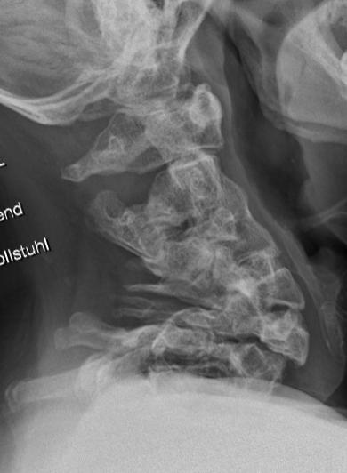
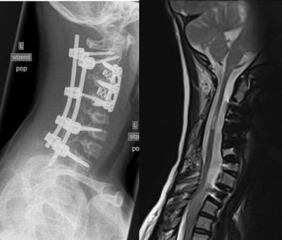
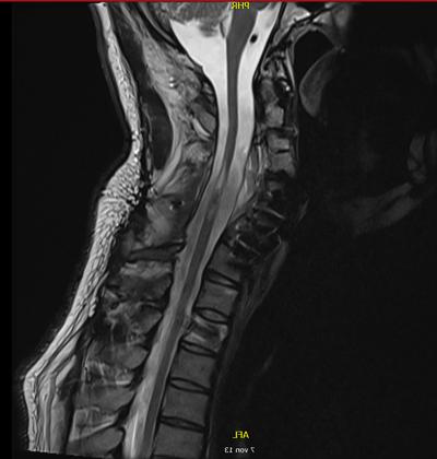
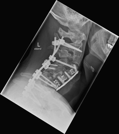
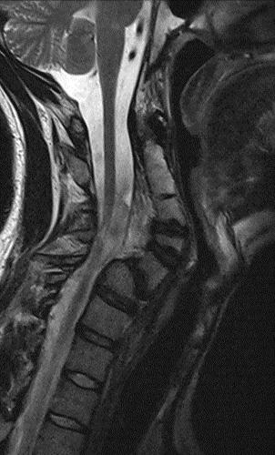
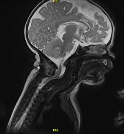
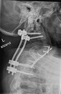
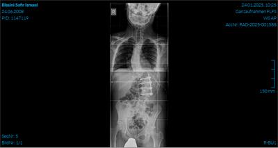
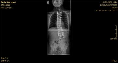
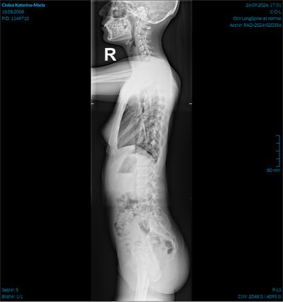
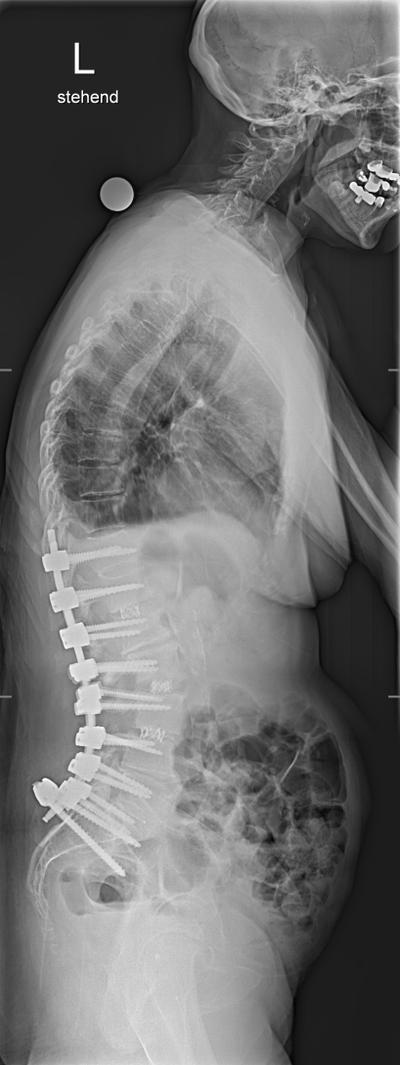
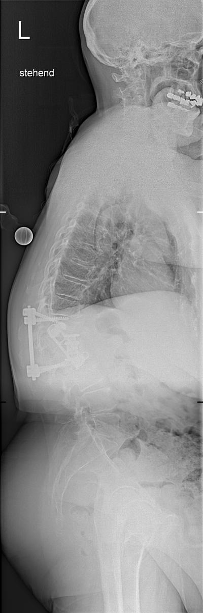
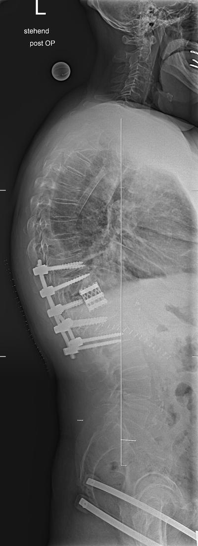
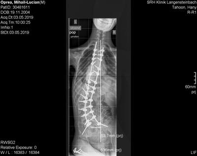
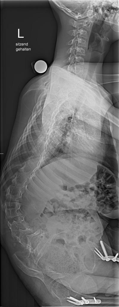
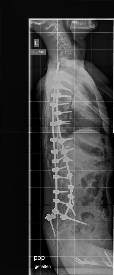
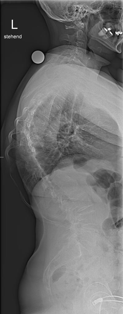
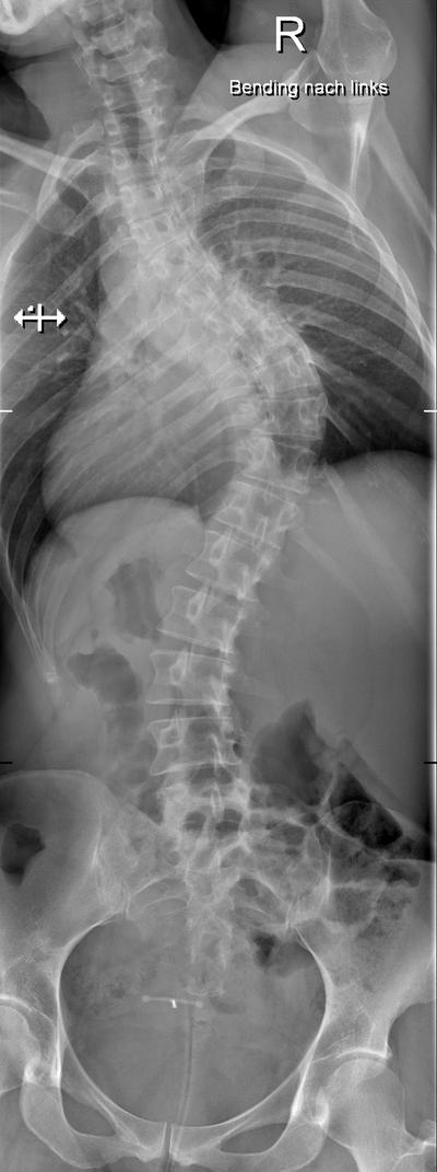
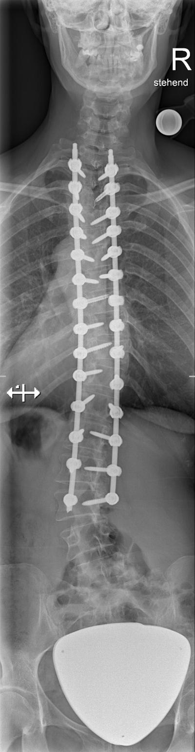
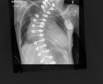
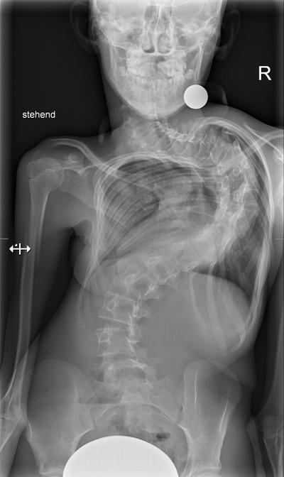
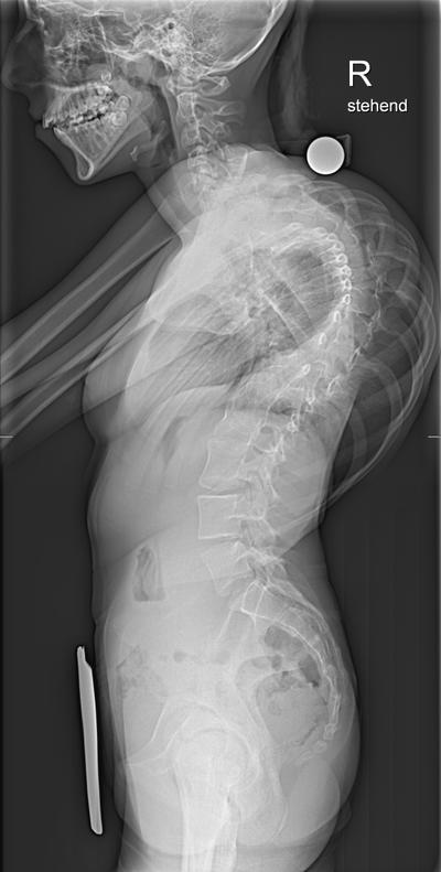
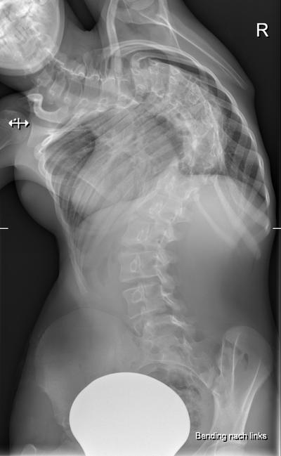
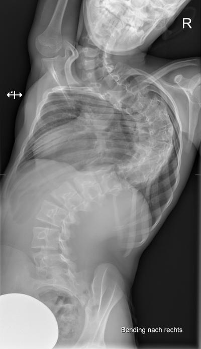
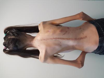
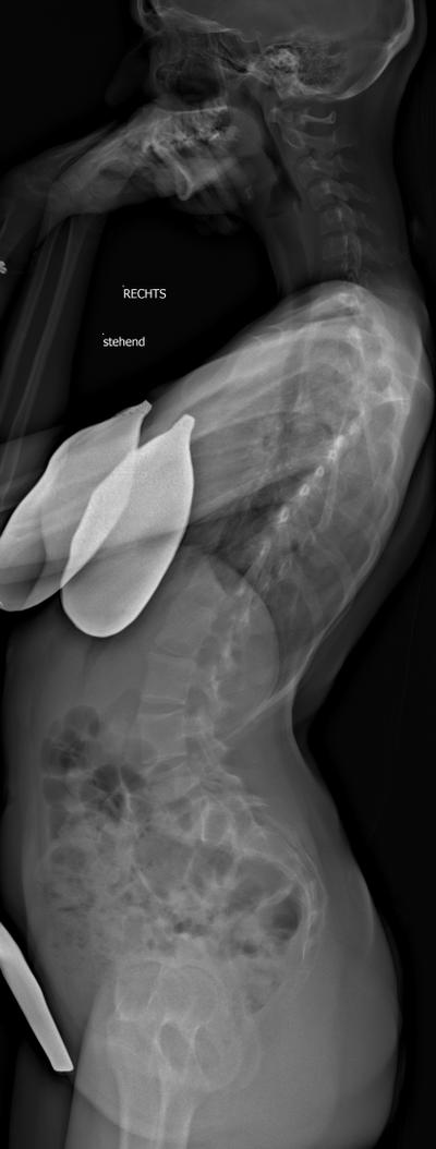
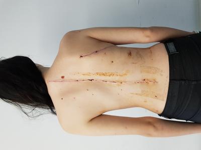
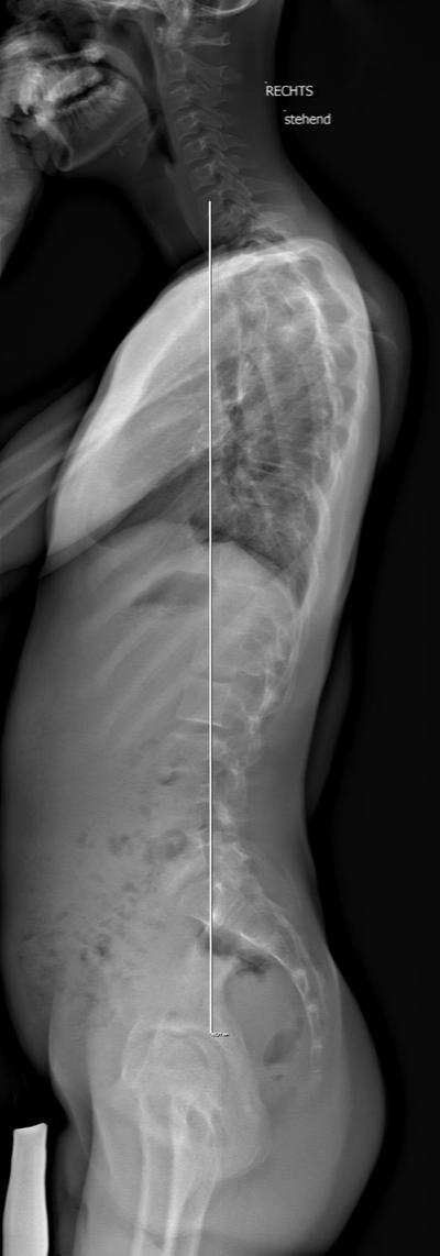
Flat Back following Posterior Correction

Slide 3

16 y.o., F, AIS Lenke 4B-.

Slide 4

Staged procedure with ventral release T6-11, halogravity traction and dorsal instrumented fusion T2-L3

After ventral release – 20°

Slide 5

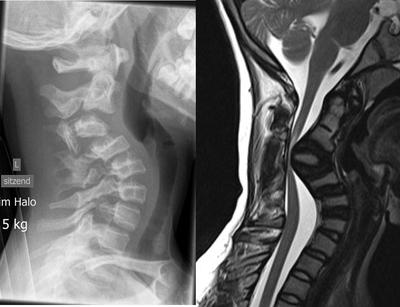
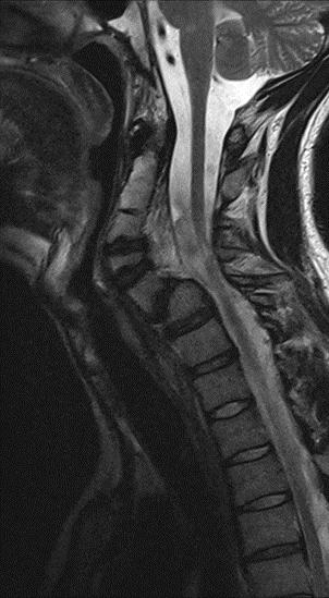
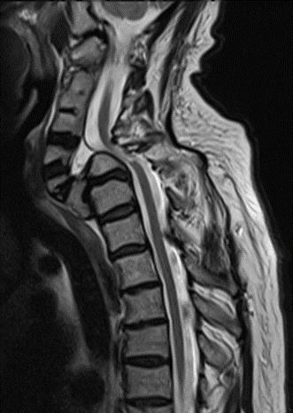
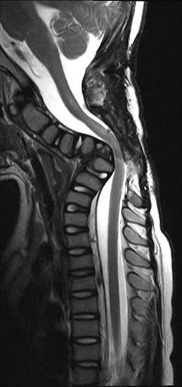
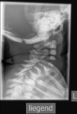
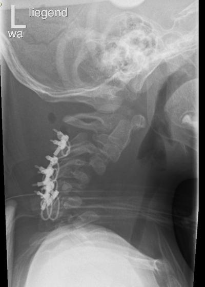
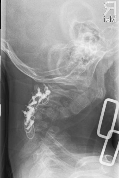
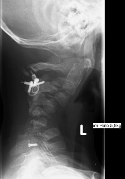
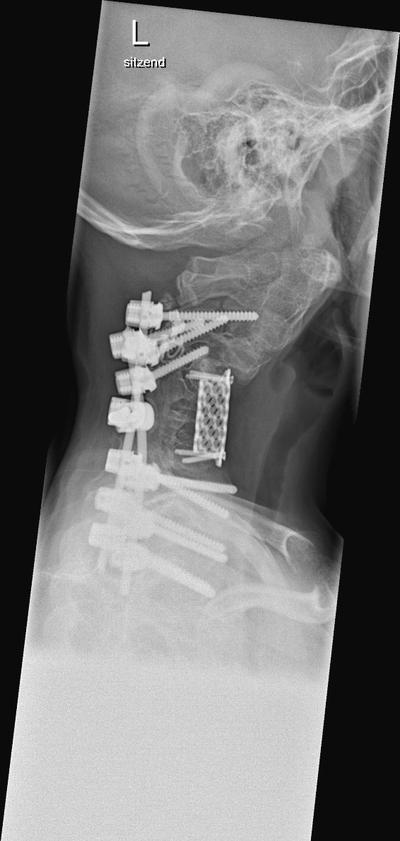
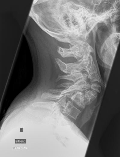
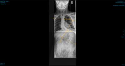
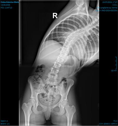
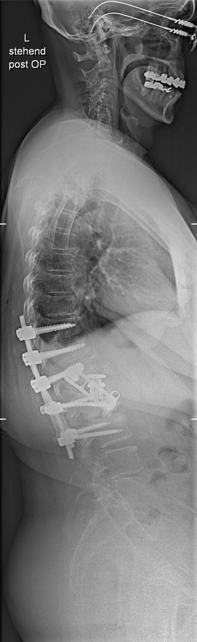
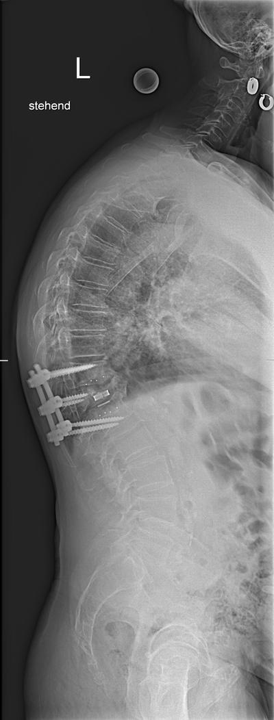
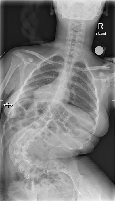
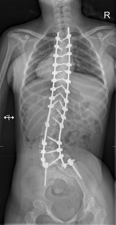
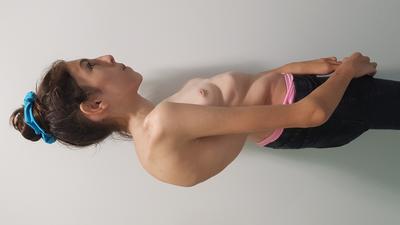
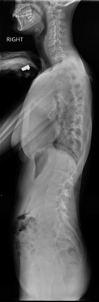
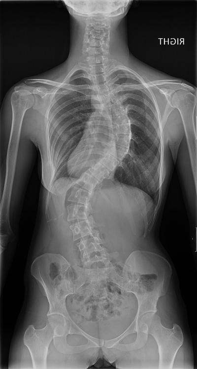
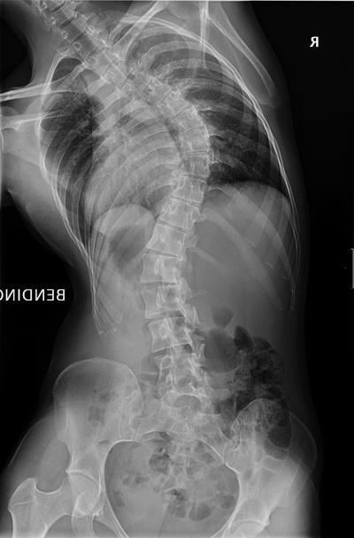
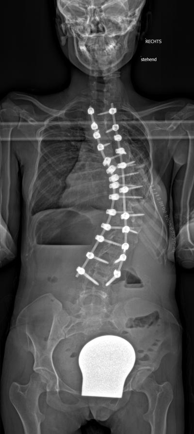
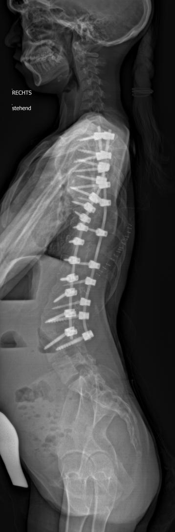
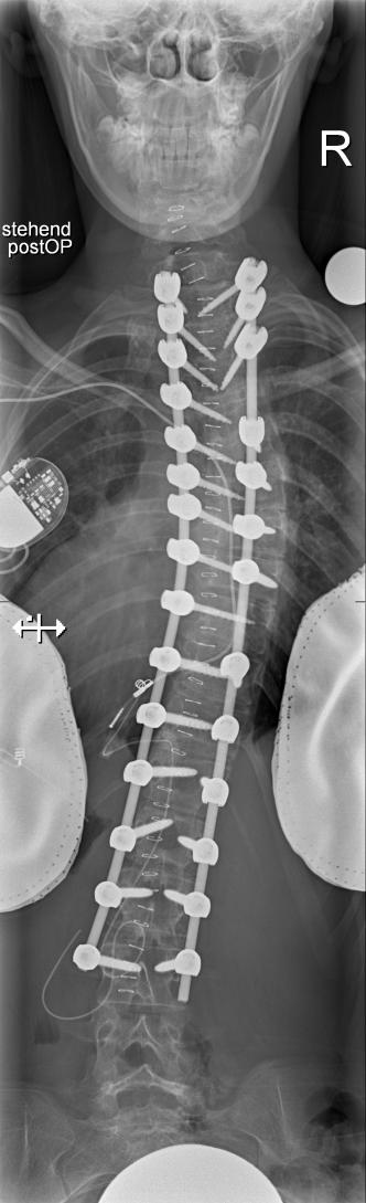
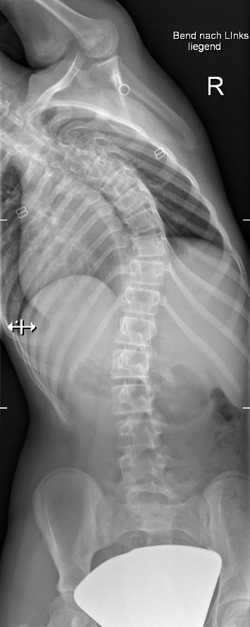
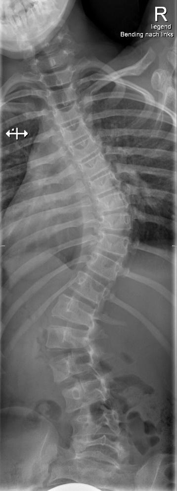
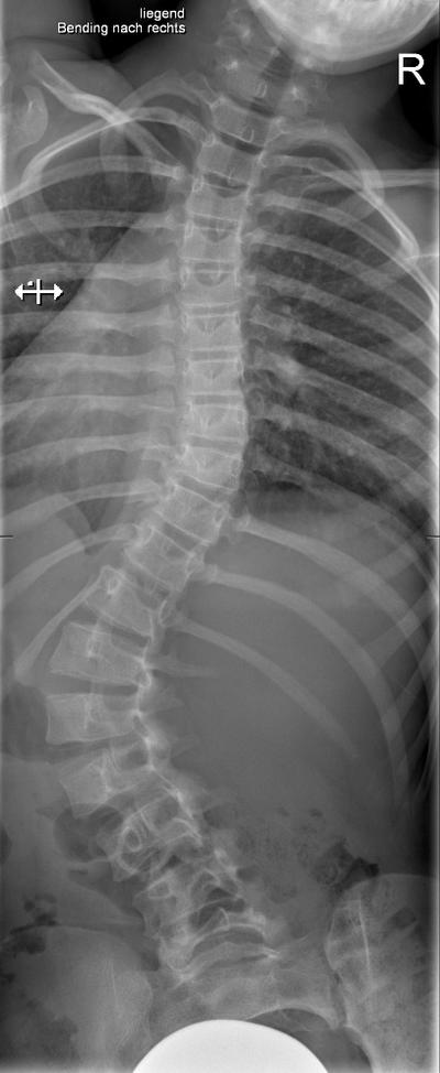
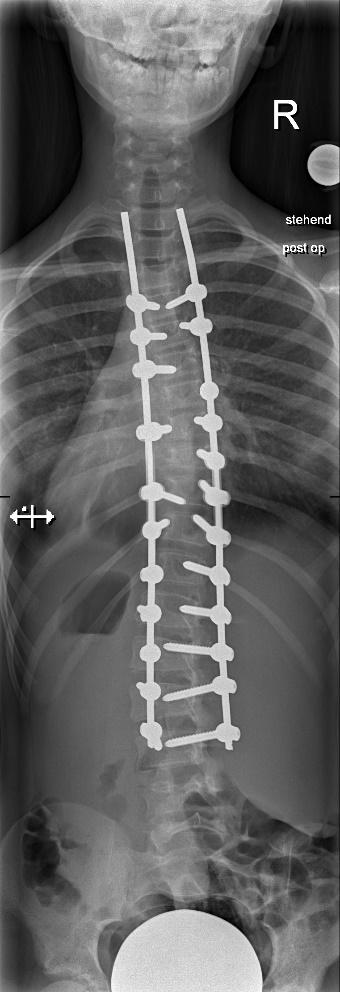
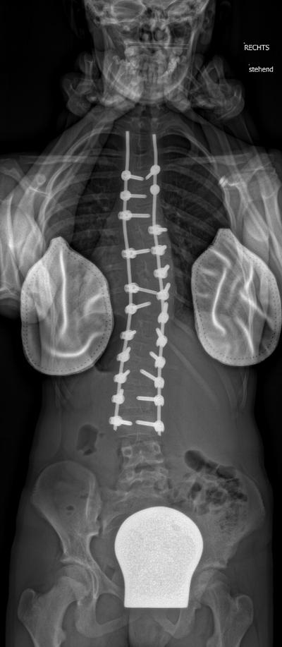
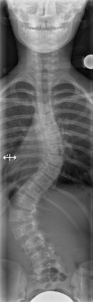
13 y.o. with severe idiopatic scoliosis Lenke 4B+.

Slide 6

Staged procedure with 1.ventral release T4-T11, halo-traction, 2.dorsal release and screw-placement, again halo traction and finally 3. instrumented fusion T1-L3

After ventral release and under Halo-traction

Slide 7

00.00.2020

SRH Unternehmen – Max Mustermann

7

Slide 8

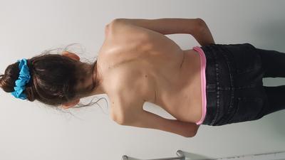
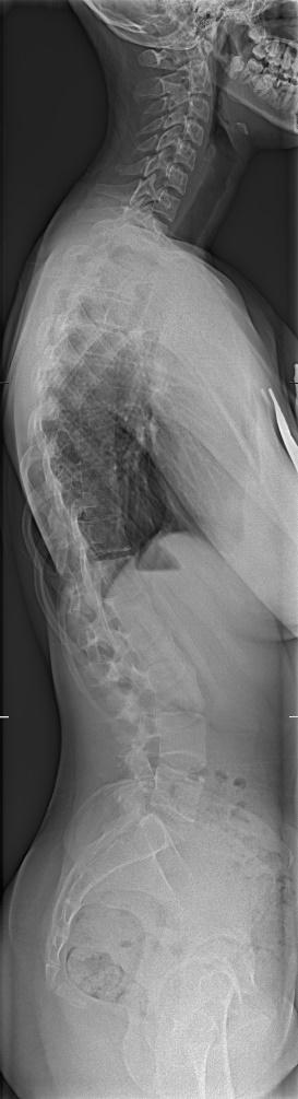
16 y.o. girl Lenke 3A-. Staged procedure with 1.ventral release T5-10 and 2.dorsal release and instrumented fusion T2-L3. Note the marked apex derotation as shown by rib re-alignment postop

Slide 9

9

00.00.2020

Slide 10

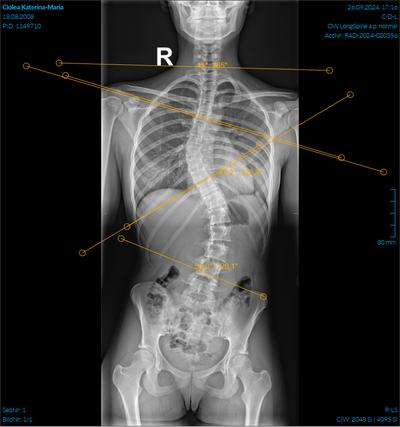
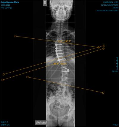
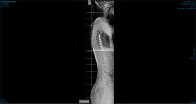
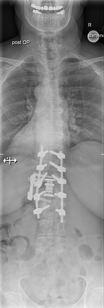
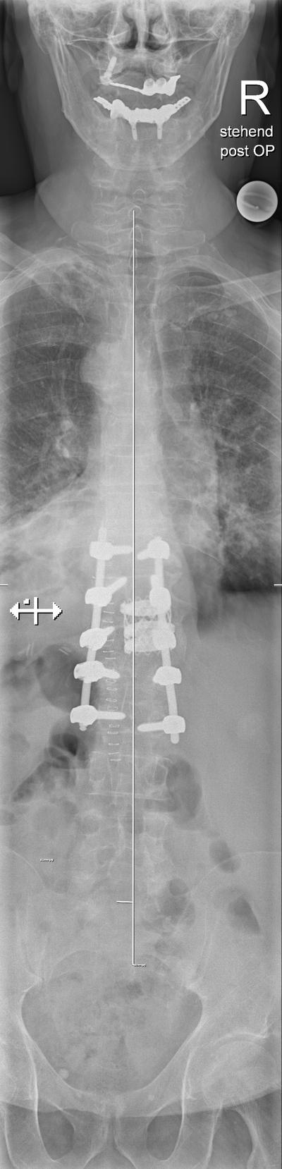
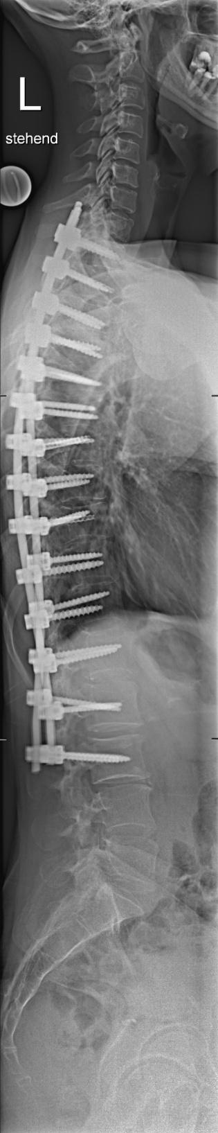
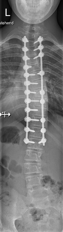
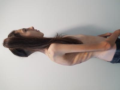
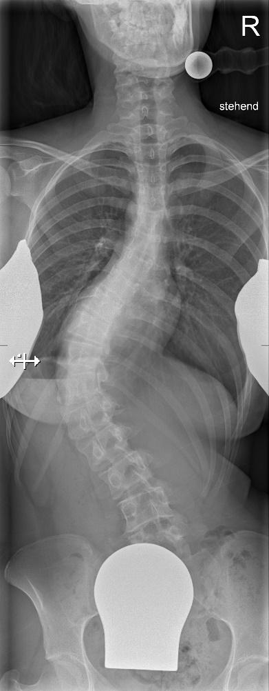
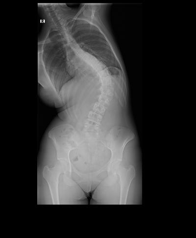
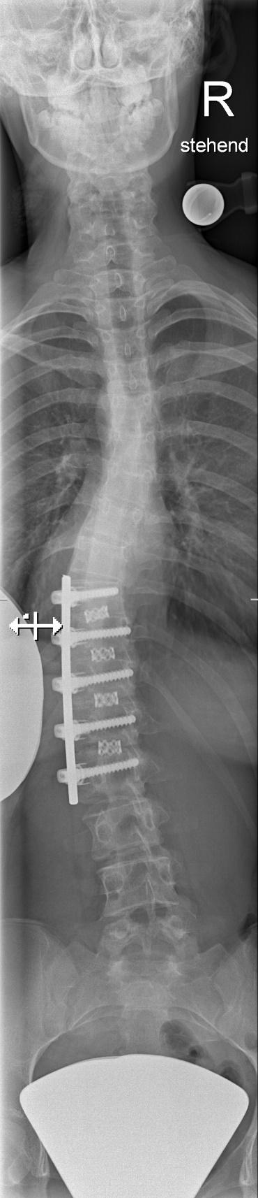
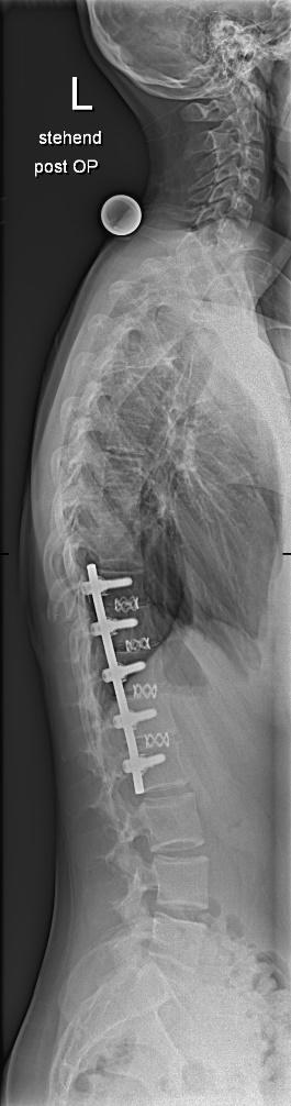
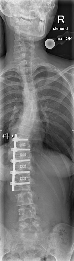
Idiopathic scoliosis Lenke 3 C- in a 28 y.o. mother.

Slide 11

Postop after1. ventral release T5-11 and 2.dorsal instrumented fusion T2-L3 in separate sessions. The LIV retained some rotation with L4 spared

After ventral release -20°

Slide 12

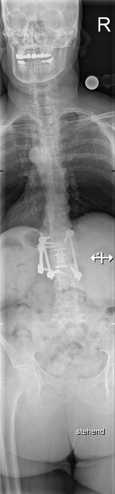
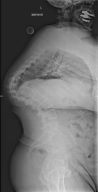
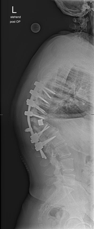
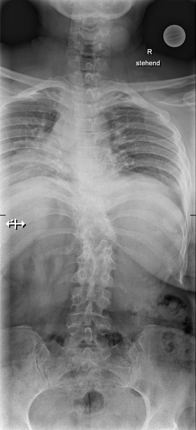
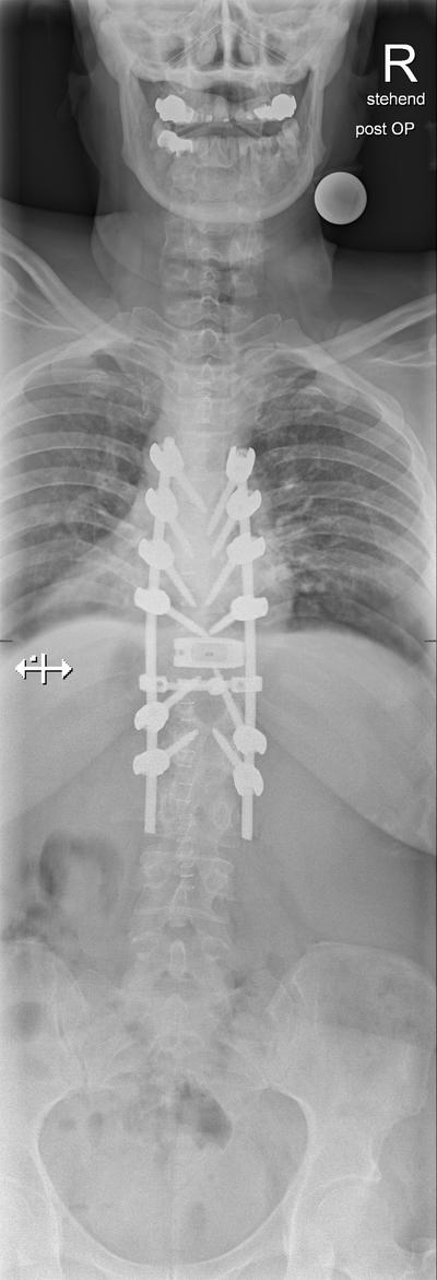
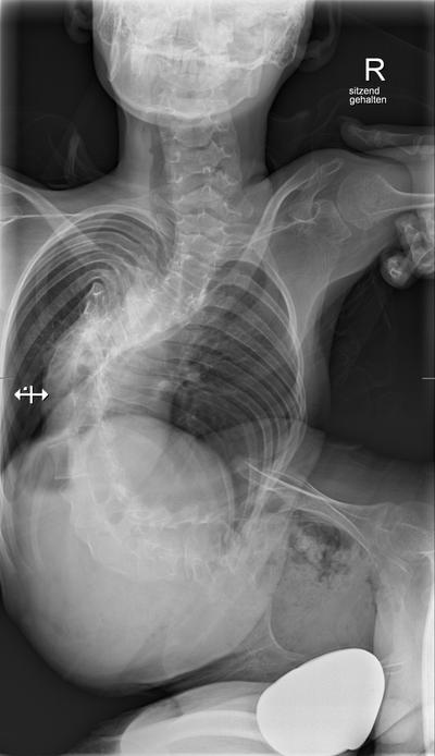
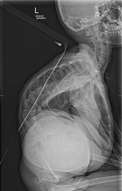
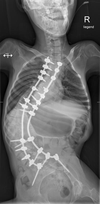
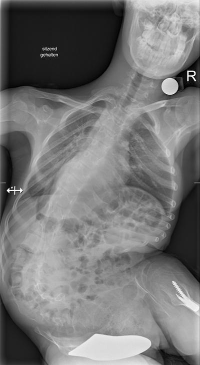
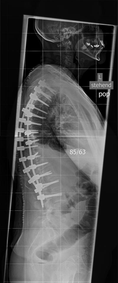
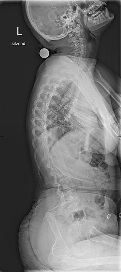
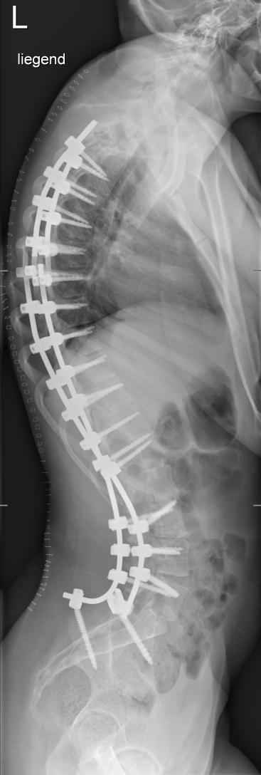
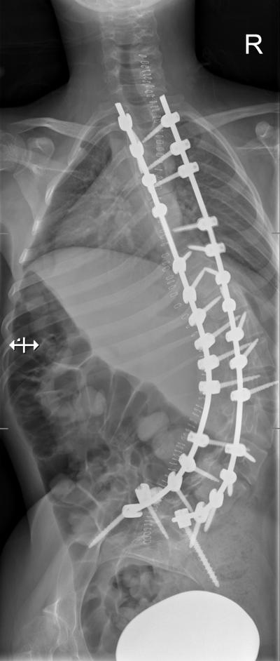
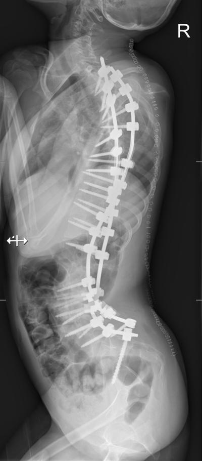
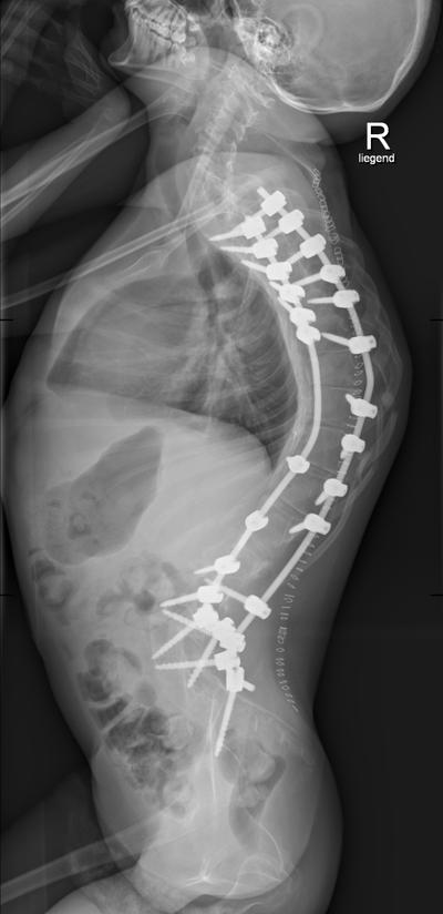
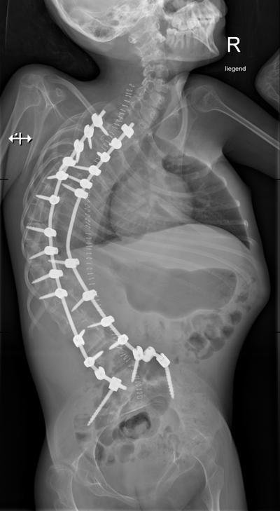
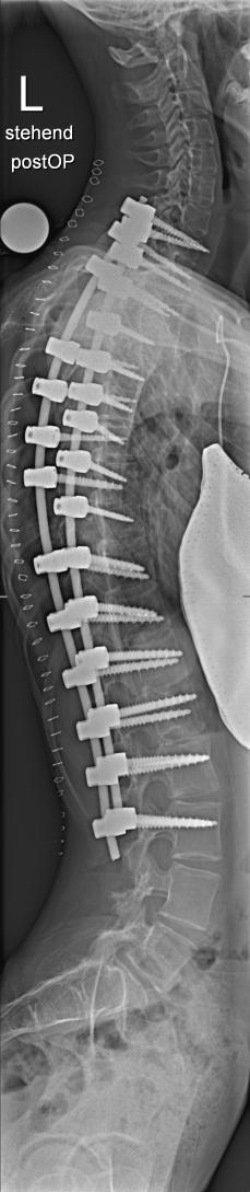
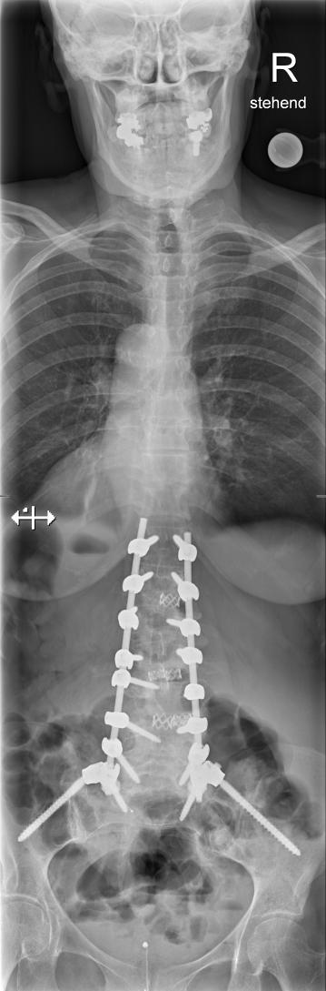
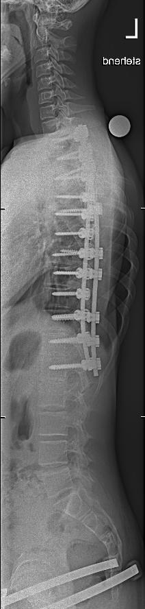
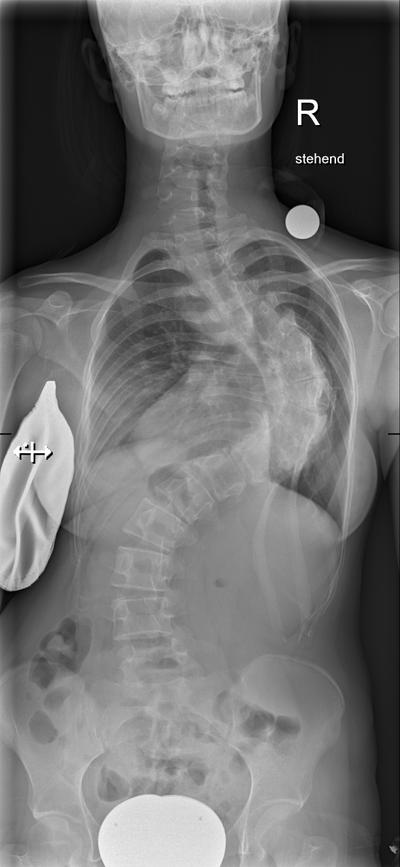
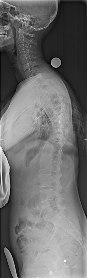
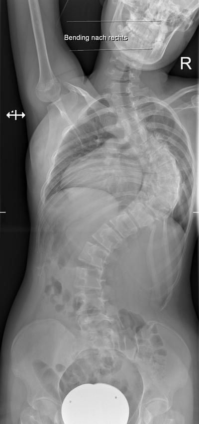
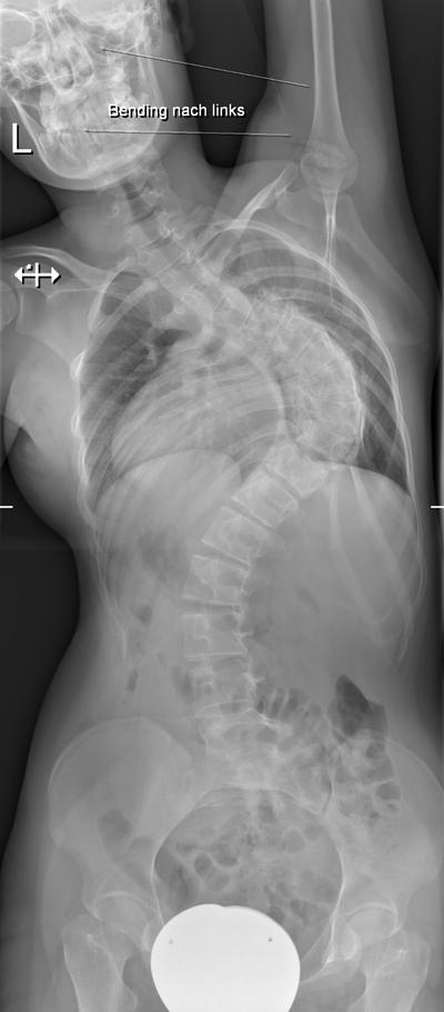
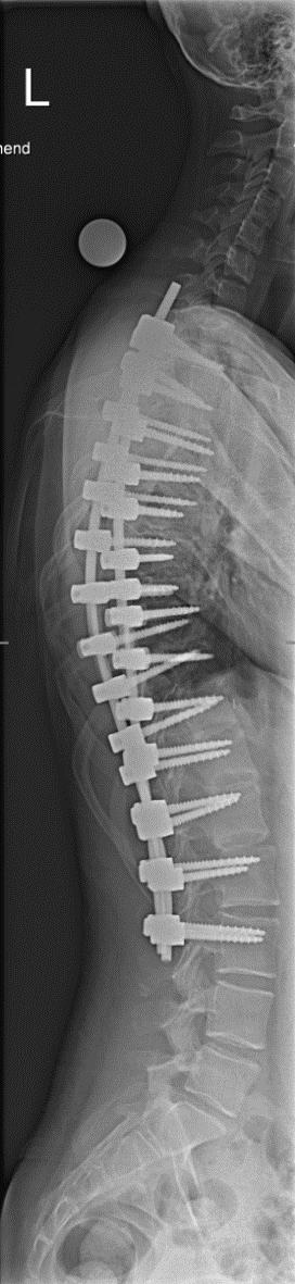
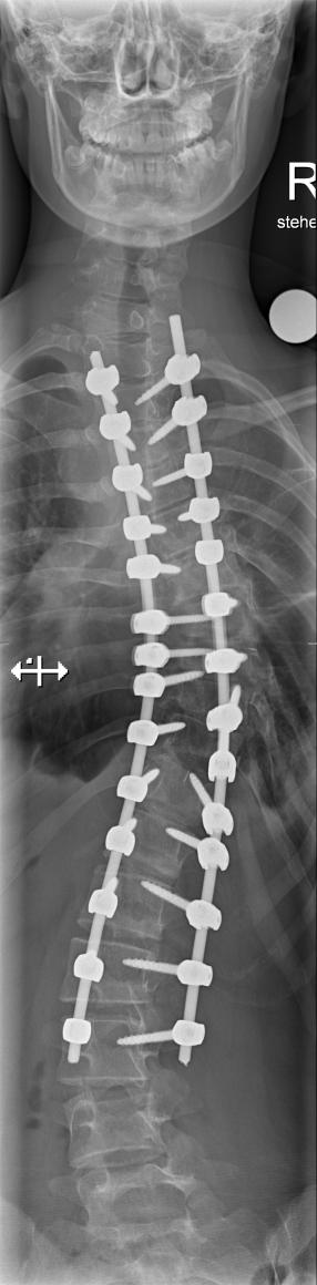
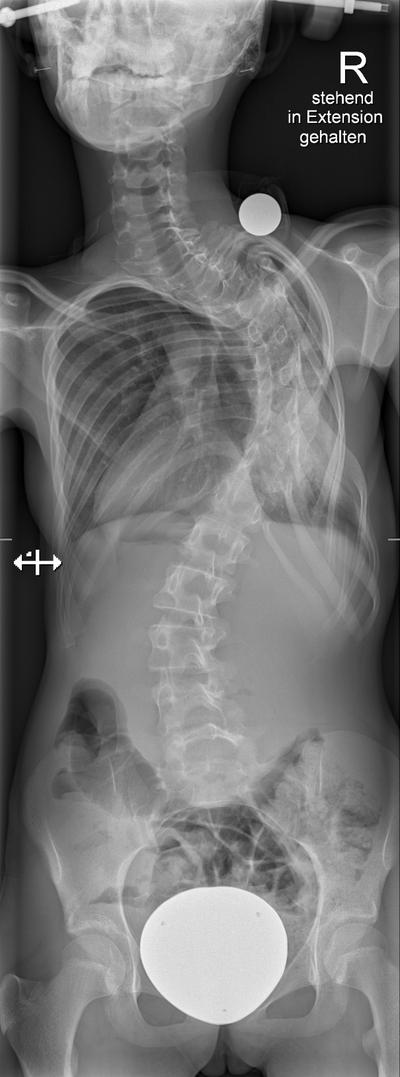
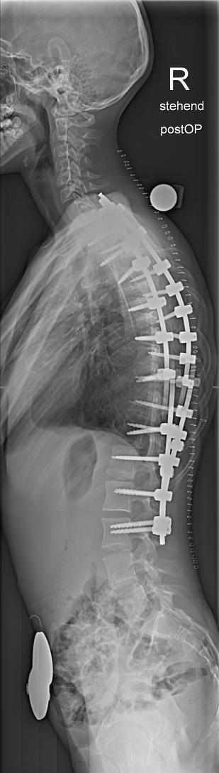
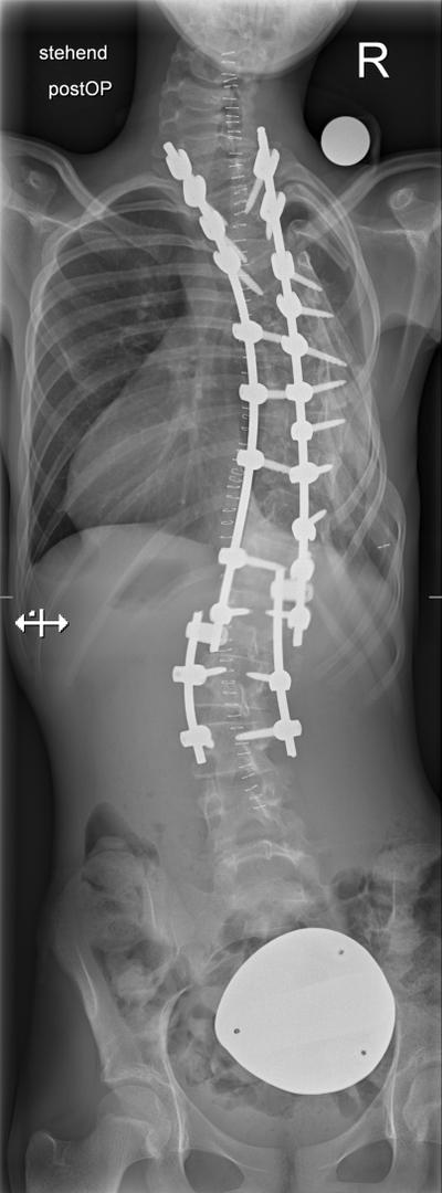
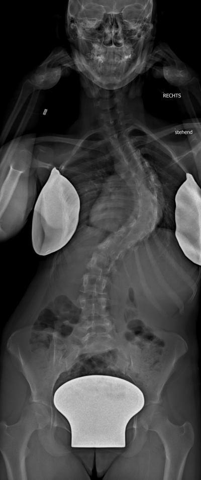
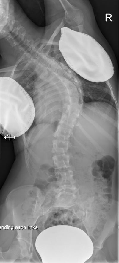
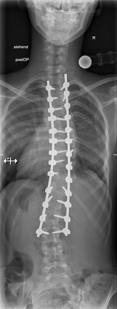
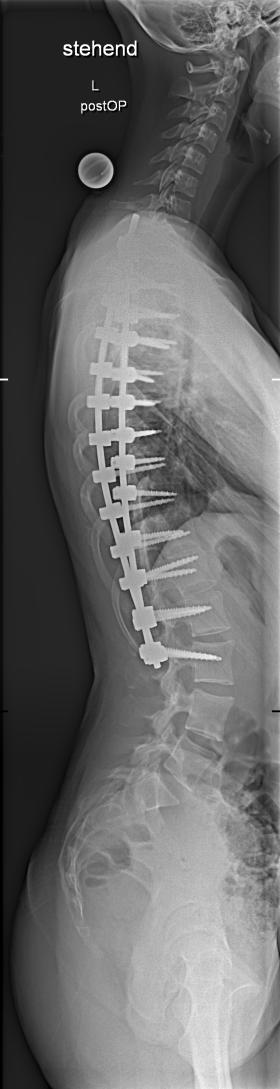
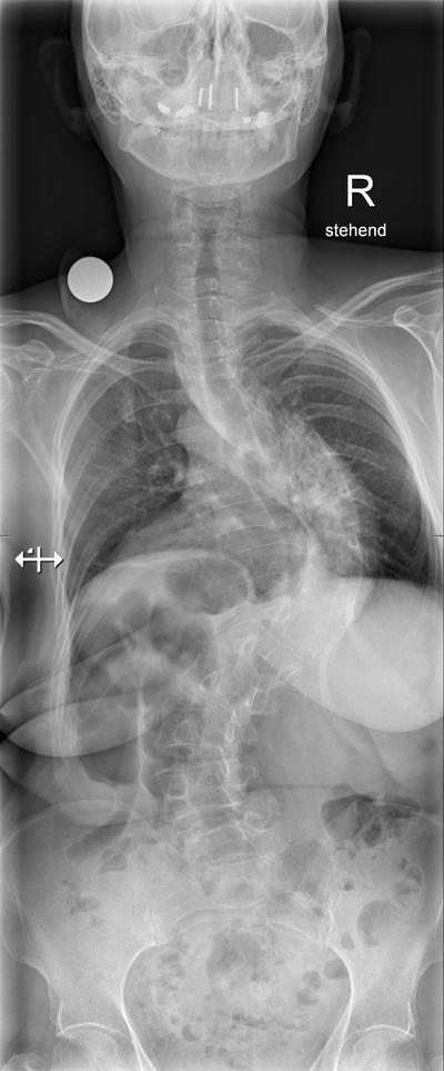
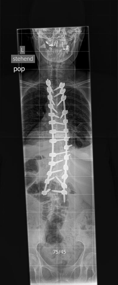
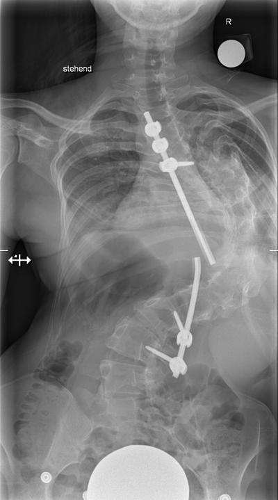
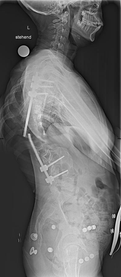
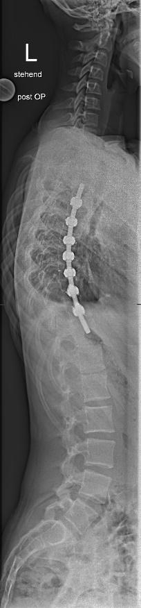
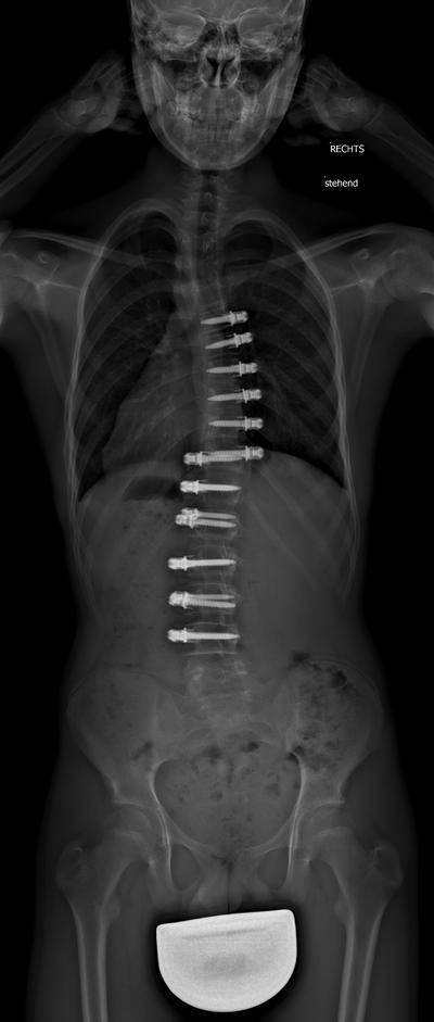
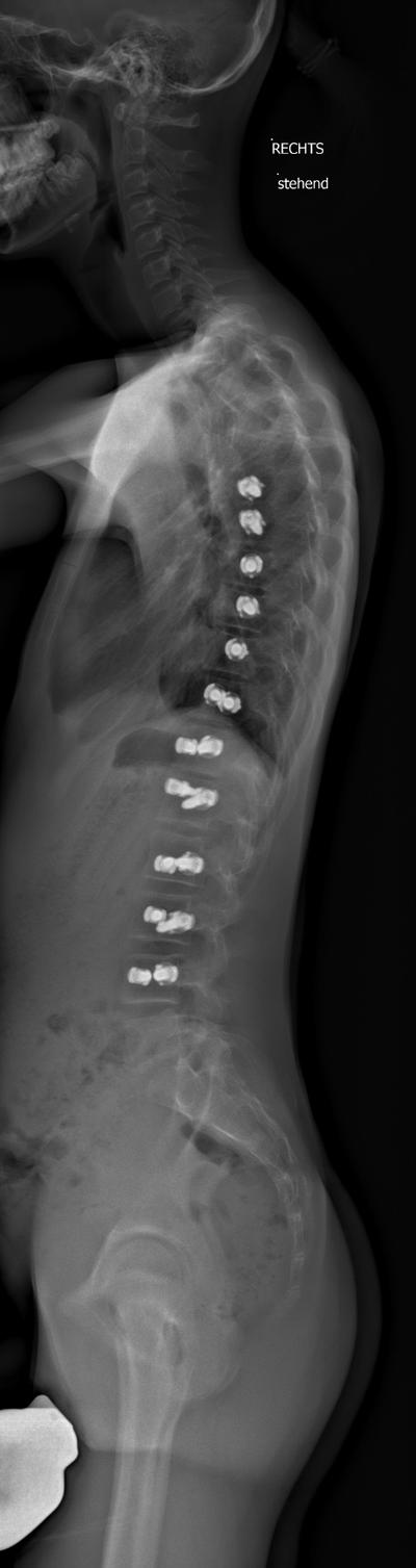
55 y.o. with AIS, stiff thoracic curve with apical spontaneous fusion. Staged approach with ventral release and subsequently dorsal instrumented fusion T2- L3

Slide 13

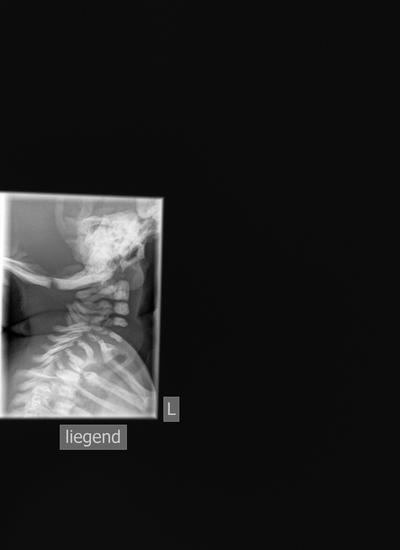
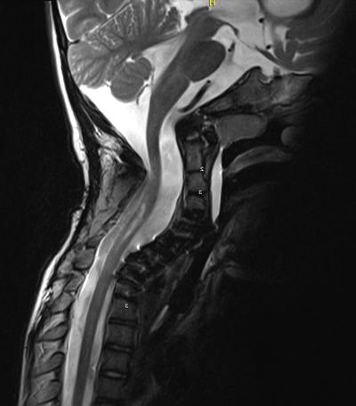
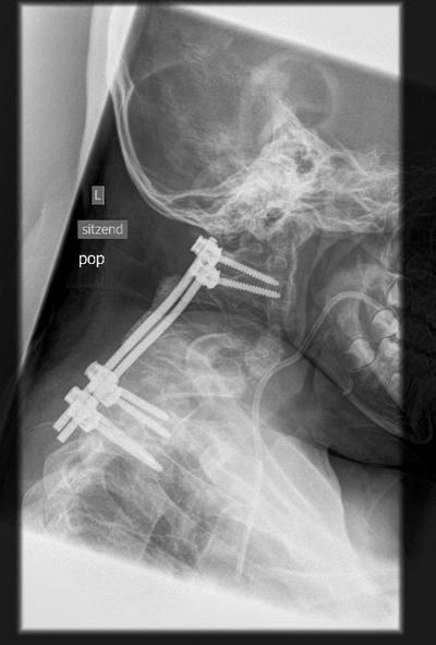
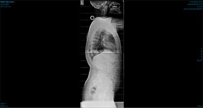
17, F, AIS, first operation elsewhere, partly fused, 1. removal of fixation and dorsal release, 2.Halotraction, 3.ventral release and 4. dorsal instrumented fusion

Slide 14

15, F, 1.dorsal release, 2.halogravity, 3.fusion. No ventral release because of impaired heart and pulmonary function.

Slide 15

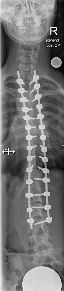
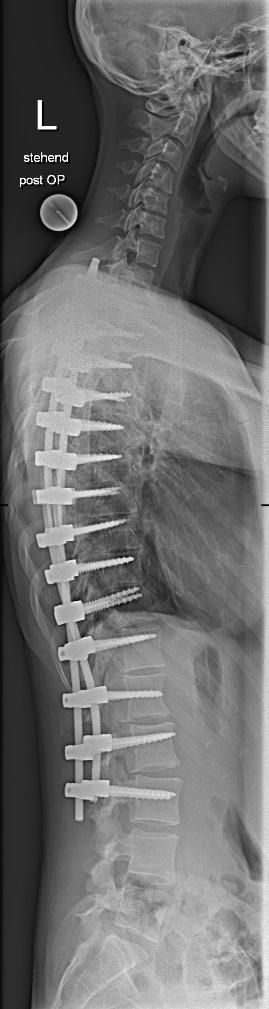
13 y.o. girl Lenke 1 B -. Should one do a ventral release and then fuse dorsally (LIV would be in the best case scenario L3) or go for a selective thoracic fusion? Ventral, dorsal or combined?

Slide 16

After ventral kyphosing derotation and selective thoracic fusion T5-T11. Postop the lumbar curve decreased from 55 to 19 degrees, we got some decent thoracic kyphosis and subsequently reduced the compensatory cervical kyphosis.

Slide 17

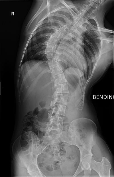
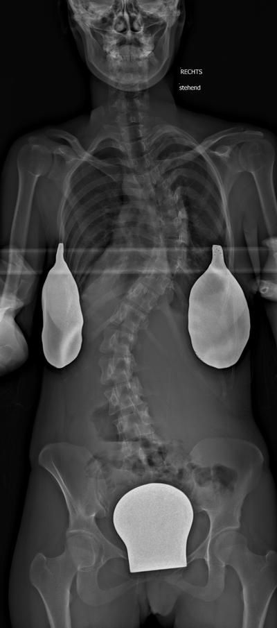
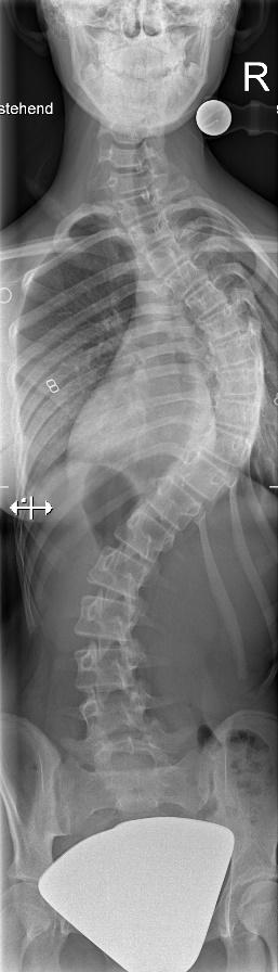
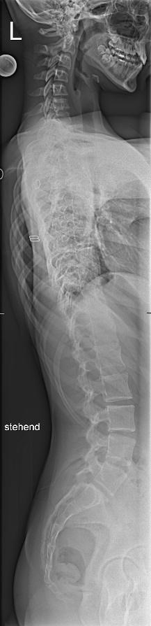
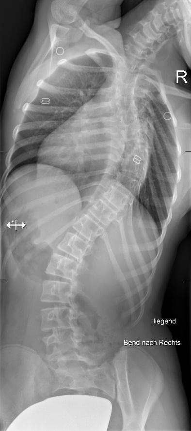
15 y.o girl AIS Lenke 1CN. Proximal thoracic and lumbar curves corect just underneath 25°. Significant rotation of lumbar vertebrae

Slide 18

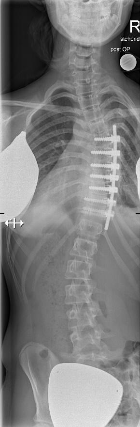
Short fusion T10-L2 through open thoracofrenolumbotomy. The lateral approach simply allows better derotation.

1 year postop

postop

Slide 19

Promises (unfulfilled?) for the futureAIS Lenke 6 in 12 y.o. Shilla growth-guidance instrumentation

1 year postop

postop

Slide 20

Promises (unfulfilled?) for the futureVertebral body tethering (VBT) – trade mobility for predictability?

Slide 21

The following posterior correction surgery is facilitated

Anterior mobilisation with shortening of the anterior column results in a substantial correction of scoliosis

A nearly complete correction in frontal as well as in sagittal plane is possible even in severe rigid deformities

The sagittal profile improves with increasing kyphosis

Correction occurs spontaneously without application of corrective forces

- Conclusions –

Combined anterior-posterior approach

Fallserie

Larsen-Syndrom — frühzeitige 360°-zervikale Fixation

14 Folien
Slide 2

Larsen syndrome: advocating early surgical treatment using 360°cervical fixation and fusion

Ioan-Lucian Branea 1, Michael Ruf, Tobias Pitzen 2, Dezsö Jeszenszky 3, Iulian Tiripa 2

1. Rhein-Neckar Wirbelsäulenzentrum, St. Josefskrankenhaus, Heidelberg, Deutschland

2. Zentrum für Wirbelsäulenchirurgie, SRH Klinikum Karlsbad, Deutschland

3. Wirbelsäulenchirurgie, Schulthess Klinik, Zürich, Schweiz

Slide 3

Larsen syndrome…

Is a rare, autosomal dominant, congenital disorder first described by Larsen et al. 1950 (defect of Filamin-B encoding gene)

Features multiple dislocation of the great joints, flat facial shape, prominent foreheads and depressed nasal bridges, hypertelorism, brachydactyly

Other features may include short stature, hypermobility, cleft palate, hearing loss

3

Slide 4

Spinal anomalies in Larsen syndrom

Occur in 84% of individuals with Larsen syndrome, 50% are in the cervical spine*

No bony connection between dorsal and anterior elements, pedicles remain cartilaginous

Consequently: olisthesis, segmental kyphosis, neurological impairment…

Knowledge of the natural history, early diagnosis and adequate therapy is paramount!

*Johnston CE, 2nd, Birch JG, Daniels JL. Cervical kyphosis in patients who have Larsen syndrome. J Bone Joint Surg Am. 1996

Slide 5

Material and methods/results

retrospective analysis of 5 consecutive patients with C-spine anomalies whithin Larsen disease treated in Langensteinbach and Zürich (2012 – 2023)

predominant focal deformity was segmental kyphosis (2) and olisthesis(3)

neurological deficit was present in all patients

all 5 patients were preoperatively unable to walk due to progressive neurological deterioration and major joints subluxation

5

Slide 6

Material and methods/results

Mean age 17 years (2- 49 years), 2 ♀, 3 ♂

All received preoperative halo-gravity traction, followed by staged procedures with back/front or front/back decompression - fixation – fusion

4 cases received primary treatment, 1 case was a revision (after dorsal laminar wiring elsewhere)

Postoperative all improved neurologically, 3 patients regained their walking ability

Complications: screw – pullout in 1, plate-breakage in 1, both revised

Slide 7

♂, 3, preoperative x-ray

postoperative after laminar wiring elsewhere

after 3 months marked kyphosis progression

after partiall removal of dorsal implants and halo-gravity traction

Slide 8

Postop x-ray after ventro-dorsal instrumented fusion C2-T4

follow-up 7 years after

Sagital CT-reconstruction pre- and postop

Slide 9

Postop MRI showing adequate cord-decompression. X-ray after removal of a loosed anterior screw.

♂, 49, preop MRI and x-ray

Slide 10

10 y.o. patient, progresive tetraparesis with loss of ambulation

Postoperative images after halo-gravity traction, staged dorso-ventral decompression and fusion C2-T1. Patient regained walking ability

Slide 11

♂, 22, progressive tetraparesis

Postoperative result, patient regained ambulation

Slide 12

♂, 2 y.o., x-ray and MRI

Postop MRI showing excellent cord decompression.

X-ray 1 year postop showing broken anterior plate

X-ray 8 year postop

Slide 13

Discussion

Patients with Larsen syndrome should be screened for segmental kyphosis of the cervical spine and early neurological deficit

Fixation and fusion of the dorsal elements alone will most likely fail in kyphosis

We recommend dorsal fixation using pedicle screws

Larsen syndrome has an early indication for 360° decompression/ fixation/ fusion

13

Slide 14

Thank you!

Fallserie

AIS Lenke 6 — anteriore derotierende Fusion

3 Folien
Slide 1

Anterior derotating fusion for AIS in Lenke 6

Finally, some time after leaving my previous workplace in Karlsbad-Langensteinbach, I have been able to resume my endeavours in adolescent scoliosis in Sankt-Josef Hospital in Heidelberg.

Ventral corection in idiopathic scoliosis has its classic indication in Lenke 1 and 5 type curves. Here are 2 interesting cases of „of the shelf“ use of anterior correction done recently:

First case - a 16 y.o. teenager with Lenke 6 curve measuring 87 degrees lumbar and 67 thoracic. He underwent a staged procedure with, at first, anterior derotating fusion of lumbar apex from T12 to L3. After the final dorsal correction operation both thoracic and lumbar curves were at 27 and 30 degrees, with a comparatively well balanced trunk.

I always dreaded inserting pedicle screws in a very rotated and laterally displaced lumbar apex such as this (especially in not skinny patients), on the other hand, from lateral, one is very close to the apical vertebral bodies.

The LIV is far from neutral, but fusing more distally would have compromised hopes of a good longterm result.

Second case – 16 y.o. girl with a Lenke 6 curve. Selective anterior fusion of the lumbar and thoracic apex led to decrease of the lumbar curve from 54 to 29 degrees and of the thoracic curve from 47 to 24 degrees by only fusing 1 (one!) lumbar and 5 thoracic segments. The „classic“ solution would have meant going dorsally from at least T4 to L3, even though a potential complete correction of the curves could have been achieved. Hybrid VBT would have been another , although also „unorthodox“, potential strategy.

Slide 2
Slide 3
Fallserie

Parkinson-Skoliose

1 Folien
Slide 1

F, 81y.o., degenerative and iatrogenic scoliosis, Parkinson disease

OP Branea 3/20

Fallserie

Thorakolumbale Kyphose — Revisionsserie

4 Folien
Slide 1

Law of the series: 3 interesting/problematic revision surgery for rigid thoracolumbar kyphosis in January 2023;

SCROLL NEXT PAGE

Slide 2

Case 1: Female, 63 y.o., failed L1 corporectomy and short (to short) instrumented percutaneous fixation in another hospital. Pseudarthrosis and painfull kyphosis. Here I used tho old cage as fulcrum and did a closing-wedge procedure through segmental piece-meal L1 spondylectomy, all dorsal. The lateral MACS –plate is in my experience very hard to revise, had to leave it in, fortunately was very loose and didn´t hindered the reposition

SEE NEXT

Slide 3

Case 2. Male, 63 y.o., failed T12 corporectomy and short (to short) instrumented percutaneous fixation in another hospital. Pseudarthrosis and painfull kyphosis. Did a re-thoraco-frenotomy, distracted and and replaced the cage using the still intact endplates on the right-side, then dorsal re-fixation and fusion

SEE NEXT

Slide 4

Case 3: Female, 53. This was very tricky; probably congenital (postraumatic or post-Pott disease also possible). I did an opening-wedge osteotomy all dorsal; I had to partially resect 6 ribs, 6 pedicles (maybe 8, I really can´t say) cut 6 roots and resect 3 (maybe 4) hypoplastic vertebral bodies, distract anteriorly, insert a fulcrum and compress dorsally. The cross-link connector is there only to protect the buckled dural sac which protruded dorsal

THANKS FOR WATCHING

Fallserie

Neuromuskuläre Skoliosen

17 Folien
Slide 1

15 y.o., neuromuscular scoliosis, paraparesis, severe pelvic obliquity. Postoperative patient regains wheelchair/bedside sitting ability

OP Branea 2019

Slide 2

15.y.o., neuromuscular scoliosis, incomplete tetraparesis. Good correction in coronal plane in spite of high curve rigidity

OP Branea 2019

Slide 3

Good sagital correction, patient can sit again on bedside and wheelchair

OP Branea 2019

Slide 4

55 y.o., f, stiff thoracic curve with apical spontaneous fusion. Staged approach with ventral release and subsequently dorsal instrumented fusion T2- L3

OP Branea 2019

Slide 5

50, F, AIS Lenke4B

Slide 6

14 y.o. girl, cerebral palsy, non-ambulatory, regains sitting ability postop

Slide 7

Also 14 y.o. , cerebral palsy, extremely rigid curve, regains sitting ability postop

Slide 8
Slide 9
Slide 10

17, F, Matex, 1.dorsal release, 2.Halotraction, 3.ventral release and dorsal fusion

Slide 11
Slide 12

15, F, 1.dorsal release, 2.halogravity, 3.fusion. No ventral release because of impaired heart and pulmonary function.

OP Branea 8/19

Slide 13

15, F, 1.dorsal release, 2.halogravity, 3.fusion. No ventral release because of impaired heart and pulmonary function.

OP Branea 8/19

Slide 14

F, 81y.o., degenerative and iatrogenic scoliosis, Parkinson disease

OP Branea 3/20

Slide 15

F, 81y.o., degenerative and iatrogenic scoliosis, Parkinson disease

OP Branea 3/20

Slide 16

F, 81y.o., degenerative and iatrogenic scoliosis, Parkinson disease

OP Branea 3/20

Slide 17

F, 81y.o., degenerative and iatrogenic scoliosis, Parkinson disease

OP Branea 3/20